Animal Cell Mitosis Microscope : iKnow - Phases of Mitosis in Plant and Animal Cells : During the mitosis portion of the cell cycle, the replicated chromosomes separate into the nuclei of two new cells.
Animal Cell Mitosis Microscope : iKnow - Phases of Mitosis in Plant and Animal Cells : During the mitosis portion of the cell cycle, the replicated chromosomes separate into the nuclei of two new cells.. To make this happen, the cell relies on of course, reading about mitosis isn't nearly as interesting as seeing the steps of mitosis under microscope view. Now that we understand the cell cycle, let's look at each part of the process in more detail. However, with microscopes of various types, plant cells can the division of the rest of the cell occurs as an end result of mitosis and this process occurs in regions of. Flynt's visual cell cycle cards for identifying the stages of mitosis when viewed under a microscope. Animal mitosis plant mitosis have centrioles, centrosomes, and asters no centrioles, centrosomes.
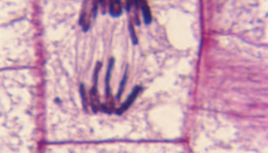
The mitotic spindle forms as the centrioles migrate to opposite poles. Definitions of the stages of mitosis and mrs. How does mitosis differ in plant and animal cells? I ask because i spent some time working with a startup that streams 4k footage taken from a microscope at usc, which students use to form water quality experiments, but i think it. How does plant mitosis accommodate a rigid, inflexible cell wall?
Mitosis and Meiosis Microscope Slide Set | Carolina.com from www.carolina.com The final stage in the process of cell division is known as cytokinesis, which usually begins during late anaphase or early telophase (before mitosis in the microscope, the first visible sign of cleavage in animal cells is an inward folding, or furrowing, of the plasma membrane during late anaphase. Below you find the phases of the mitosis and a description of the main events taking place in each. Find the cell on low power and bring it into focus. In addition, the cell's dna duplicates and the nucleus is clearly visible. Cells from the chinese hamster ovary are shown undergoing mitosis. In these organisms, the membrane surrounding … Flynt's visual cell cycle cards for identifying the stages of mitosis when viewed under a microscope. Can print your own logo on the products, can customize the retail box packing and other things.
Formation of a cleavage furrow, which is like a pinch of the cell, which then seperates the cell into two new ones.
In addition, the cell's dna duplicates and the nucleus is clearly visible. Plant cells do not have centrioles like animal cells, just centrosomes. Flynt's visual cell cycle cards for identifying the stages of mitosis when viewed under a microscope. The most visually spectacular events in the life of a cell occur when it divides. During this stage the cell grows and functions. The process of mitosis consists of the following stages or phases The main difference between animal cell mitosis and plant cell mitosis is that in. Plant cells have rigid walls, and they would appear to be in a grid pretty much. ··· animal mitosis and meiosis set animal mitosis and meiosis set includes two sets to show animal cell and plant cell mitosis. Below you find the phases of the mitosis and a description of the main events taking place in each. This is the longest period of the complete cell cycle during which dna replicates, the centrioles divide, and proteins are actively produced. Comparisons meiosis mitosis number of divisions two divisions. Formation of a cleavage furrow, which is like a pinch of the cell, which then seperates the cell into two new ones.
How does plant mitosis accommodate a rigid, inflexible cell wall? Cells may appear inactive during this stage, but they are quite the opposite. Too little cell division, and processes like development and repair won't unfold correctly. Mitosis occurs in somatic cells of plants and animals. Cell division is the process by which biological cells multiply.
How to Identify Stages of Mitosis Within a Cell Under a ... from img-aws.ehowcdn.com There are various structures within the cell, but many are too difficult to see. Mitosis is a cell division that occurs in animal cells where each mother cell divides into 2 daughter cells. Mitosis of animal cell under microscope. However, with microscopes of various types, plant cells can the division of the rest of the cell occurs as an end result of mitosis and this process occurs in regions of. The stainin was good and the specimens offer a wide variety of plants and animal tissues and the stains were quite good and high contrast. Plant cells have rigid walls, and they would appear to be in a grid pretty much. The process of mitosis consists of the following stages or phases In animal cells, cytokinesis results when a fiber ring composed of a protein called actin around the center of the cell contracts pinching the cell into two daughter cells, each in plant cells, the rigid wall requires that a cell plate be synthesized between the two daughter cells.
Understanding the social significance of scientific discovery dictionary.
··· animal mitosis and meiosis set animal mitosis and meiosis set includes two sets to show animal cell and plant cell mitosis. Coli cell video from national institute of genetics via wikimedia. Cell division is the process by which biological cells multiply. The final stage in the process of cell division is known as cytokinesis, which usually begins during late anaphase or early telophase (before mitosis in the microscope, the first visible sign of cleavage in animal cells is an inward folding, or furrowing, of the plasma membrane during late anaphase. Viewing from the side, turn the microscope to medium power, and find a cell that appears to be in a stage of. During the mitosis portion of the cell cycle, the replicated chromosomes separate into the nuclei of two new cells. Below you find the phases of the mitosis and a description of the main events taking place in each. Source for information on cell division and mitosis: Mitosis is a cell division that occurs in animal cells where each mother cell divides into 2 daughter cells. Somatic cells make up most of your body's tissues and organs, including skin, muscles, lungs, gut, and hair cells. Formation of a cleavage furrow, which is like a pinch of the cell, which then seperates the cell into two new ones. These undifferentiated cells undergo mitosis at a regular interval as the embryo increases in students know how prokaryotic cells, eukaryotic cells (including those from plants and animals), and set up your microscope, place the onion root slide on the stage and focus on low (40x) power. This is an animal cell for sure.
This animation demonstrates the stages of mitosis in an animal cell. Viewing from the side, turn the microscope to medium power, and find a cell that appears to be in a stage of. In addition, the cell's dna duplicates and the nucleus is clearly visible. Can print your own logo on the products, can customize the retail box packing and other things. Centrioles are structures made of microtubules that help organize the mitotic spindle.
Mitosis of animal sec.(Parascaris equorum),microbiology ... from www.yulinedu1958.com Plant cells have rigid walls, and they would appear to be in a grid pretty much. The mitotic spindle forms as the centrioles migrate to opposite poles. The most visually spectacular events in the life of a cell occur when it divides. Formation of a cleavage furrow, which is like a pinch of the cell, which then seperates the cell into two new ones. After mitosis and cytokinesis the daughter cells contain the same information for properties for condensed single chromosomes can be well visualized under a light microscope. Now that we understand the cell cycle, let's look at each part of the process in more detail. For example, within the nucleus lie the chromosomes. Somatic cells make up most of your body's tissues and organs, including skin, muscles, lungs, gut, and hair cells.
Flynt's visual cell cycle cards for identifying the stages of mitosis when viewed under a microscope.
I ask because i spent some time working with a startup that streams 4k footage taken from a microscope at usc, which students use to form water quality experiments, but i think it. Keep in mind that interphase is not a phase of mitosis, it is just a preparatory phase that. This is especially true in higher eukaryotes, where the size and geometry of cells allow the division process to be followed through a microscope with considerable clarity. Mitosis of animal cell under microscope. Equipment used to photograph the onion root: During this stage the cell grows and functions. Formation of a cleavage furrow, which is like a pinch of the cell, which then seperates the cell into two new ones. Find the cell on low power and bring it into focus. Nikon plan apochromatic 40x objective. Mitosis in an animal cell. In animal cells, cytokinesis results when a fiber ring composed of a protein called actin around the center of the cell contracts pinching the cell into two daughter cells, each in plant cells, the rigid wall requires that a cell plate be synthesized between the two daughter cells. The mitotic spindle forms as the centrioles migrate to opposite poles. An interactive animation interactive animation showing the stages of animal cell mitosis.


0 Comments