Diagram Of Animal Cell Seen Under Electron Microscope : A Labeled Diagram Of The Animal Cell And Its Organelles Biology Wise : Cell membrane dr jastrow s electron microscopic atlas.
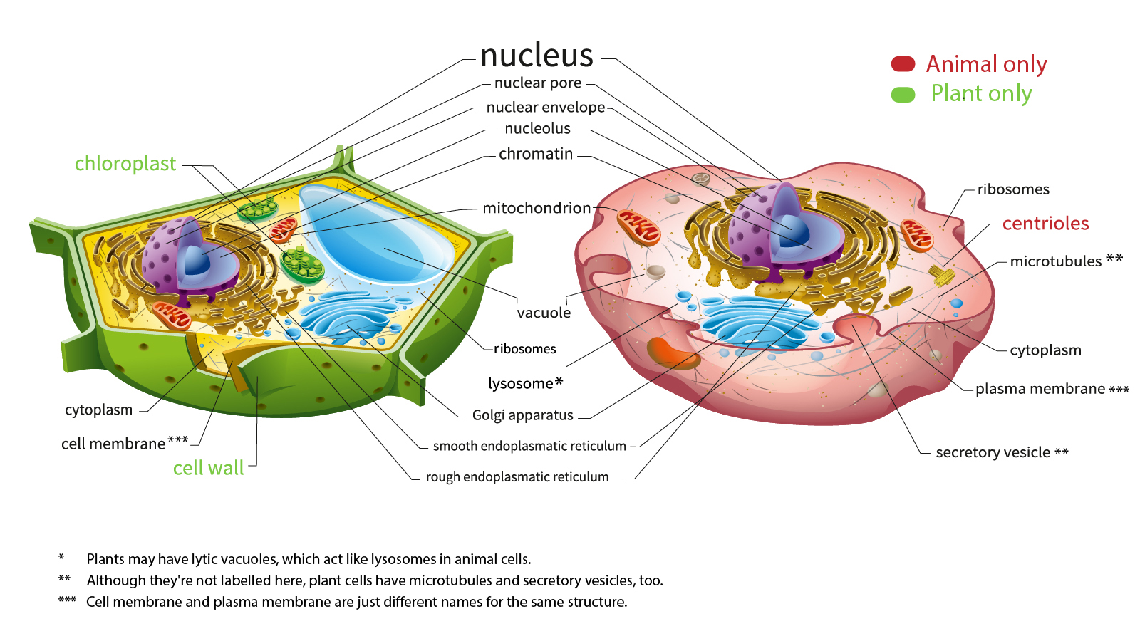
Diagram Of Animal Cell Seen Under Electron Microscope : A Labeled Diagram Of The Animal Cell And Its Organelles Biology Wise : Cell membrane dr jastrow s electron microscopic atlas.. Study the two diagrams of plant and animal cells below. The diagram below shows a specified plant cell(solved). Here's a photo of a plant cell under an electron microscope. Image:animal cell seen under electron microscope. Most cells, both animal and plant, range in size between 1 and 100 micrometers and are thus visible only with the aid of a microscope.
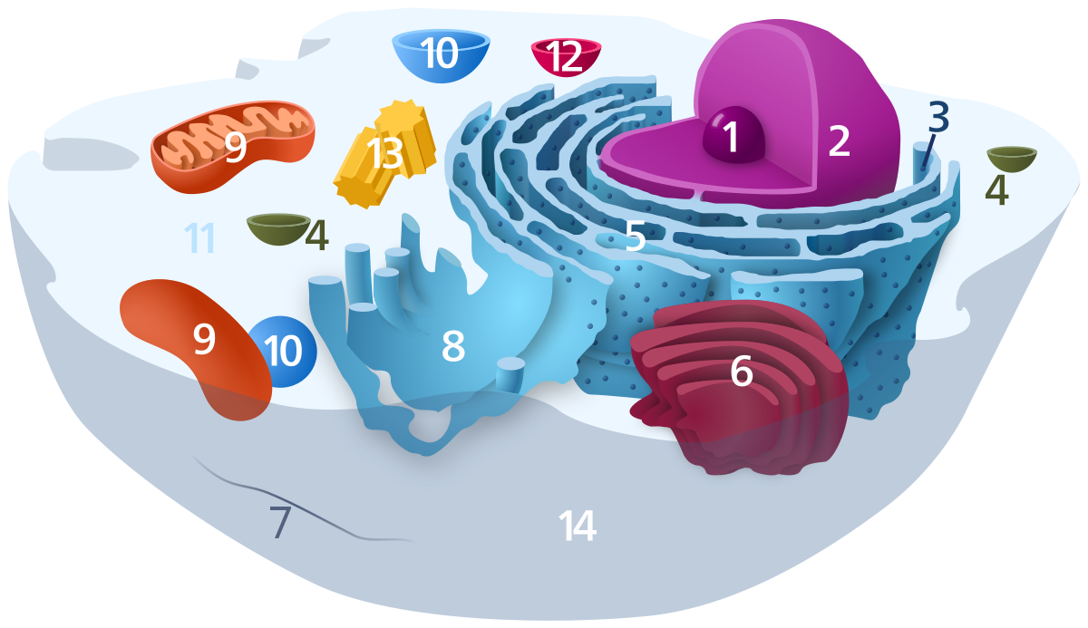
Given below is the diagram of a cell as seen under the microscope after having been placed in a solution Each type is specialised to do a particular role. However, when you use an electron microscope to increase the magnification many thousands of times you see that these seemingly simple structures are incredibly complex, each with its own specialized function. A cell is a very tiny structure which exists in living bodies. Typical animal cell pinocytotic vesicle lysosome golgi vesicles golgi vesicles rough er (endoplasmic reticulum) smooth er download this awesome diagram.
Ribosome Wikipedia from upload.wikimedia.org Light and electron microscopes allow us to see inside cells. We say cells are microscopic because they can only be seen under a microscope. Electron microscopes use electron beams focused by electromagnets to magnify and resolve microscopic specimens. The diagram shows part of a cell surface membrane. The diagram is taken from an electron micrograph of a cell. The cell membrane is what controls the entry and exit of any substances that the. 3 the electron microscope two types transmission electron microscope (tem) scanning electron microscope (sem) activity read 8 ultrastructure of a plant cell as seen through an electron microscope. Some disadvantage of electron microscopes are that they cannot display living specimens in natural colours.
(ii) presence of large central vacuole in plant cell.
Cell is a tiny structure and functional unit of a living organism containing various parts known as organelles. The animal cell is more fluid or elastic or malleable in structure; Lysosomes are the cell's garbage disposal. Animal cells have another set of organelles not found in plant cells: After this, add another oval shape outside the line you just drew, and this will make the cell membrane to your animal cell. How is it different from animal cell? Electron microscopes use electron beams focused by electromagnets to magnify and resolve microscopic specimens. Light and electron microscopes allow us to see inside cells. A.robert hooke:studied cork section and name the. 3 the electron microscope two types transmission electron microscope (tem) scanning electron microscope (sem) activity read 8 ultrastructure of a plant cell as seen through an electron microscope. Rana ray diagram of animal cell seen through electron. Removing cellular waste products from the cell. Illustrate only a plant cell as seen under electron.
Bring your presentation to life. Illustrate only a plant cell as seen under electron. As for seeing electrons under any microscope in general, i would say we have come as close to it as scientifically and technically possible the diagram is very clear, and labeled; The diagram is taken from an electron micrograph of a cell. Here's a photo of a plant cell under an electron microscope.
Plant Cell Diagram Electron Microscope The Greatest Garden Plant Cell Diagram Cell Diagram Animal Cell Structure from i.pinimg.com A) what is the formula for calculating linear magnification of a specimen when using a hand lens b) give a reason why staining is necessary when preparing specimens for observation under the microscope. Bring your presentation to life. Removing cellular waste products from the cell. University holds stem cell research boon wee looked through a microscope to study the structure of a cell. It also has a very high resolving power. Under the microscope, an animal cell shows many different parts called organelles, that work together to keep the cell functional. Rabies, seen here under a microscope, is an often fatal viral disease that a generalised animal cell as observed under an electron microscope. Rana ray diagram of animal cell seen through electron.
Animal and plant cell under electron microscope.
These ensure that the organism functions as a whole. Given below is the diagram of a cell as seen under the microscope after having been placed in a solution As for seeing electrons under any microscope in general, i would say we have come as close to it as scientifically and technically possible the diagram is very clear, and labeled; University holds stem cell research boon wee looked through a microscope to study the structure of a cell. Animal and plant cell under electron microscope. Cell scanning electron microscope hd stock video 717 725 243. Ishita observed a slide of eukaryotic cell under electron microscope. The plant cell as more rigid and stiff walls. Now the first thing to point out when looking at images under an electron microscope is the scale. But at the same time it is interpretive. Light and electron microscopes allow us to see inside cells. Cell structure i nucleus medical media. However, when you use an electron microscope to increase the magnification many thousands of times you see that these seemingly simple structures are incredibly complex, each with its own specialized function.
The diagram below shows a specified plant cell(solved). Animal cell (as seen under electron microscope). Rana ray diagram of animal cell seen through electron. Animal cells have another set of organelles not found in plant cells: See how a generalized structure of an animal cell and plant cell look with labeled diagrams.
Here S How Plant And Animal Cells Are Different Howstuffworks from cdn.hswstatic.com Lysosomes are the cell's garbage disposal. Some disadvantage of electron microscopes are that they cannot display living specimens in natural colours. 9 pupil activity cell structure read through the information on each of the organelles as. The diagram shows a stage micrometer, with divisions 23. The diagram shows part of a cell surface membrane. Cell structure and organisation_notes igcsebiology dnl. Which represents the same cell, seen under a light (optical) microscope at 400 magnification? Be the first to comment.
Cell structure and organisation_notes igcsebiology dnl.
Animal and plant cell under electron microscope. 9 pupil activity cell structure read through the information on each of the organelles as. We say cells are microscopic because they can only be seen under a microscope. Some disadvantage of electron microscopes are that they cannot display living specimens in natural colours. Cell membrane dr jastrow s electron microscopic atlas. A) what is the formula for calculating linear magnification of a specimen when using a hand lens b) give a reason why staining is necessary when preparing specimens for observation under the microscope. There are many different types of cells in animals. Plant animal cells staining lab answers schoolworkhelper. Ishita observed a slide of eukaryotic cell under electron microscope. But at the same time it is interpretive. Cell scanning electron microscope hd stock video 717 725 243. It is the outermost membrane of an animal cell having a thickness. Here is the microscopic view of animal cell.


0 Comments