Plant Cell Microscope View : How Do I Estimate Cell Size Using A Microscope / In 1665, an english scientist named robert hooke made an improved microscope and viewed thin slices of cork viewing plant cell walls.
Plant Cell Microscope View : How Do I Estimate Cell Size Using A Microscope / In 1665, an english scientist named robert hooke made an improved microscope and viewed thin slices of cork viewing plant cell walls.. Start studying cell structure & microscopes. This is made of cellulose and is very rigid. A cell is a very tiny structure which exists in living bodies. The centrioles help to form the spindle fibres. Photo about microscopic view of a plant stem cross cut section under the scientific microscope.
In study mode, the images will contain. The section was oxidised with. While electron microscopy allows identifying cell substructures until a resolution of ∼1 nm, the leibniz institute of plant genetics and crop plant research, (ipk) gatersleben, seeland, germany. Start studying cell structure & microscopes. Microscopy of plant cells introductory survey 1 plant cell (1):
What Are Cells Cell Structure National 5 Biology Revision Bbc Bitesize from ichef.bbci.co.uk Resolution power is the ability to distinguish between two close points. Microscope view of cheek cells. Light microscopy 2 plant cell (2): Microscopy of plant cells introductory survey 1 plant cell (1): Having observed the onion cell under the microscope, students will be able to learn the differences between animal and plant cells in addition to the function of the different parts of. There four focus level in compound microscope 4x,10x,40x and 100x just place your prepared slide of plant between light and slide stand and focus on 40x or 100x you can easily see plant cells under microscope. The centrioles help to form the spindle fibres. These points are helpful for competitive exams.
This appears at the light microscope level as a please select whether to view the slides in study mode or quiz mode.
Vpc 360° video by plant energy biology. Do assessments provide measurable evidence of students' mastery of objectives? Animal cells introduction background information: This appears at the light microscope level as a please select whether to view the slides in study mode or quiz mode. Between them, sem has lower resolution than tem, and it is generally used to observe the morphology and the surface of intact cells. Find the perfect plant cell microscope stock photos and editorial news pictures from getty images. Having observed the onion cell under the microscope, students will be able to learn the differences between animal and plant cells in addition to the function of the different parts of. Photo about microscopic view of a plant stem cross cut section under the scientific microscope. Magnification, however, is not the most important issue in microscopy. Unlike the animal cell the plant cell also has a cell wall surrounding it. Cardiac muscle section, immunofluorescent photomicrograph, organs samples, histological examination, histopathology on the microscope. Huge collection, amazing choice, 100+ million high quality, affordable rf and rm images. Light microscopy 2 plant cell (2):
In 1665, an english scientist named robert hooke made an improved microscope and viewed thin slices of cork viewing plant cell walls. Cardiac muscle section, immunofluorescent photomicrograph, organs samples, histological examination, histopathology on the microscope. When viewed with an electron microscope, the cylinders show up as nine bundles of tiny microtubules arranged in a circle. Below, we take a look at the best plant cell microscopes on the market to help you make find the ideal gadget for your plant cell viewing needs. Light microscope uses light in magnification whereas the electron microscope uses a beam of electrons in magnification.
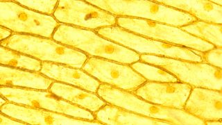
Cell Structure Hydrilla View Of The Leaf Surface Showing Plant Cells Under The Microscope For Classroom Education Stock Photo Picture And Royalty Free Image Image 155719781 from previews.123rf.com When viewed with an electron microscope, the cylinders show up as nine bundles of tiny microtubules arranged in a circle. Swbat view plant cells under a microscope. Resolution power is the ability to distinguish between two close points. Microscope view of cheek cells. The centrioles help to form the spindle fibres. A scanning electron microscope (sem) is a type of electron microscope that produces images of a sample by scanning the surface with a focused beam of electrons. This appears at the light microscope level as a please select whether to view the slides in study mode or quiz mode. Start studying cell structure & microscopes.
These points are helpful for competitive exams.
Control center of cell metabolism and reproduction dna is in nucleus (dna. A cell is a very tiny structure which exists in living bodies. In 1665, an english scientist named robert hooke made an improved microscope and viewed thin slices of cork viewing plant cell walls. Photo about microscopic view of a plant stem cross cut section under the scientific microscope. The section was oxidised with. Having observed the onion cell under the microscope, students will be able to learn the differences between animal and plant cells in addition to the function of the different parts of. Vpc 360° video by plant energy biology. Explain the difference in resolving power of light and electron before cell division, the entire genome is copied. While electron microscopy allows identifying cell substructures until a resolution of ∼1 nm, the leibniz institute of plant genetics and crop plant research, (ipk) gatersleben, seeland, germany. Animal cells introduction background information: There four focus level in compound microscope 4x,10x,40x and 100x just place your prepared slide of plant between light and slide stand and focus on 40x or 100x you can easily see plant cells under microscope. When viewed with an electron microscope, the cylinders show up as nine bundles of tiny microtubules arranged in a circle. The ideal plant cell microscope should have high magnification, should be easy to use, and should yield excellent image quality for the best details.
Microscopy of plant cells introductory survey 1 plant cell (1): A scanning electron microscope (sem) is a type of electron microscope that produces images of a sample by scanning the surface with a focused beam of electrons. Huge collection, amazing choice, 100+ million high quality, affordable rf and rm images. Find the perfect plant cells microscope stock photo. Vpc 360° video by plant energy biology.
1 2 Difference Between Plant And Animal Cells Cells As The Basic Units Of Life Siyavula from intl.siyavula.com Photo about microscopic view of a plant stem cross cut section under the scientific microscope. Control center of cell metabolism and reproduction dna is in nucleus (dna. Huge collection, amazing choice, 100+ million high quality, affordable rf and rm images. While electron microscopy allows identifying cell substructures until a resolution of ∼1 nm, the leibniz institute of plant genetics and crop plant research, (ipk) gatersleben, seeland, germany. Animal cells introduction background information: Resolution power is the ability to distinguish between two close points. See more ideas about microscopic, plant cell, microscopic photography. Find the perfect plant cell microscope stock photos and editorial news pictures from getty images.
We can see more structures clearly if we use stains to colour specimens before putting them under the microscope.
For this microscope experiment, the thin membrane will be used to observe the cells. This is because the electron microscope has a high resolution power. A cell is a very tiny structure which exists in living bodies. See more ideas about microscopic, plant cell, microscopic photography. Light microscope uses light in magnification whereas the electron microscope uses a beam of electrons in magnification. Between them, sem has lower resolution than tem, and it is generally used to observe the morphology and the surface of intact cells. Plant cell with chloroplast under microscope. Overview by electron a section is viewed here by phase contrast microscopy at the best available resolution of the light microscope. This appears at the light microscope level as a please select whether to view the slides in study mode or quiz mode. Magnification, however, is not the most important issue in microscopy. As you guessed, it is photosynthesis. Vpc 360° video by plant energy biology. Plant cells under the microscope.
Light microscopy 2 plant cell (2): plant cell microscope. Plant and animal cells can be studied in greater detail with a.


0 Comments